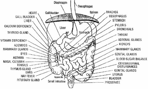
41 colon diagram with labels
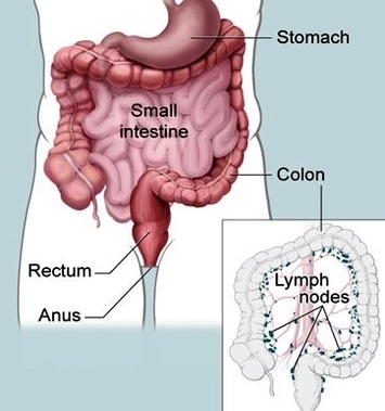
Anatomy of Colon and Rectum | SEER Training The entire colon is about 5 feet (150 cm) long, and is divided into five major segments. The rectum is the last anatomic segment before the anus. The ascending and descending colon are supported by peritoneal folds called mesentery. The right colon consists of the cecum, ascending colon, hepatic flexure and the right half of the transverse colon. Small Intestine and Large Intestine (With Diagram) - Biology Discussion 2. Propulsion: The chyme is moved over the large area of small intestine to facilitate digestion and absorption and the residues are propelled downwards to the ileocecal junction to reach the large intestine, mostly for excretion. In man, the time taken for the food to travel in the small intestine as well as in stomach can be easily estimated ...
Colon Anatomy - Medical Art Library The colon is divided into four parts: the ascending, transverse, descendingand sigmoid. The ascending and transverse colon meet at the right hepatic flexure(near the liver). The transverse and descending colon meet at the left splenic flexure(near the spleen). The large intestine has three bands of longitudinal muscle fibers called taeniae coli.
Colon diagram with labels
Sigmoid colon - Definition, Anatomy and Function | Kenhub Sigmoid colon - ventral view The gastrointestinal system is divided into the foregut, midgut and hindgut. The foregut stretches from the oesophagus to the major duodenal papilla, the midgut from the major duodenal papilla to two thirds of the transverse colon, and the hindgut from this point to the pectinate line of the rectum. Neurovasculature Abdomen and digestive system anatomy: diagrams labeled - IMAIOS Colon: three diagrams of the face of the colon and rectum, with or without the mesentery of the colon and taenia coli as well as a detailed illustration of the right colonic flexure. Large intestine Colon Anatomy (with Small Intestine Label) - National Cancer Institute 720x602. View. Download. Title: Colon Anatomy (with Small Intestine Label) Description: Drawing shows the cecum, ascending colon, transverse colon, descending colon, sigmoid colon, rectum, and anal canal. Also shown is the small intestine. The cecum connects the small intestine to the colon.
Colon diagram with labels. 40 Colon diagram Vector Images, Colon diagram Illustrations - Depositphotos 40 Colon diagram Stock Vector Images, Royalty-free Colon diagram Drawings & Illustrations. VectorMine Crohns disease vector illustration. Labeled diagram with diagnosis. VectorMine Ulcerative colitis vector illustration. Labeled anatomical infographic. Picture of the Human Colon Anatomy & Common Colon Conditions - WebMD The ileum (last part of the small intestine) connects to the cecum (first part of the colon) in the lower right abdomen. The rest of the colon is divided into four parts: • The ascending colon... Cecum Histology Slide with Labeled Image and Diagram The tunica muscular layer of the provided cecum labeled image shows two distinct smooth muscle layers - inner longitudinal or oblique bundles and outer wavy bundles. Again, the cecum images show some elastic fibers in this layer. In addition, the cecum labeled image shows a thin and loose connective tissue layer with numerous blood vessels. How does the bowel work? Bowel information with diagrams Diagram of the position and sections of the small bowel The colon The colon is divided into 4 sections. Ascending colon The first part of the colon is joined to the small bowel and goes up the right side of the abdomen (tummy). Transverse colon The second section goes across the abdomen from your right to your left side. Descending colon
Illustration Picture of Anatomy - Colon - eMedicineHealth The colon is the largest part of the large intestine, extending from the cecum to the rectum. It is 5 feet long and its function is to reabsorb water from digested food and concentrate solid waste material, known as stool. The colon is made of several sections. The ascending colon travels up the right side of the abdomen. Understanding the Human Stomach Anatomy With Labeled Diagrams Given below is a labeled diagram of the stomach to help you understand stomach anatomy. The stomach is divided into four parts. These include: Cardia Fundus Body Pylorus Cardia refers to the section of the stomach that is located around the cardiac orifice. The lower esophageal sphincter lies at the junction where the esophagus meets the stomach. Colon Histology Slide with Labeled Diagram - AnatomyLearner Colon Histology Slide with Labeled Diagram 04/06/2022 04/06/2022 by anatomylearner The colon histology slide possesses the typical four layers of a tubular organ - mucosa, submucosa, muscularis, and serosa. But, there are no permanent plica circularis and villi in the colon slide as found in the different segments of the small intestine. PDF Digestive System Diagram - direzionedidattica-vignola.edu.it Diagram . Large Intestine Mechanical Digestion Chemical Digestion Saliva Hydrochloric Acid Pepsin Trypsin Bile Lipase Stomach Small Intestine Enzymes from Liver and Pancreas Large Intestine (Transverse Colon) Descending Colon System Circulatory Kidneys #1 #2 Water and Vitamins Nutrients The Digestive System Esophagus Mouth On your Digestive ...
How do the intestines work? - Medical News Today The small intestine is roughly 20-25 feet in length, making it the longer section. It has a very high surface area, which is amplified roughly 60-120 times. The small intestine is not flat or ... anatomy and physiology colon Colon diagram with labels PPT - Basic Science - "Large Bowel" PowerPoint Presentation, free. 14 Pics about PPT - Basic Science - "Large Bowel" PowerPoint Presentation, free : Print A&p 2 Test 4 Digestive System flashcards | Easy Notecards, Another diagram of the colon On CureZone Image Gallery and also Organization of the Visceral ... Rectum & Anus | Function & Parts | Rectum Diagram Video | MyGiHealth A person uses their pelvic oor muscles and the anal sphincter to control when stools are pushed out. The puborectalis muscle is a loop of muscle that wraps around the lower rectum. This muscle relaxes and allows the rectum to straighten. The anal sphincter and pelvic floor muscles also relax. This all happens at the same time to allow stool to ... Colon Diagram High Res Illustrations - Getty Images Browse 210 colon diagram stock illustrations and vector graphics available royalty-free, ... human digestive system, with labels. - colon diagram stock illustrations. medical illustration of human torso with outlines of internal organs, side view - 19th century - colon diagram stock illustrations ...
colon lg | Anatomy System - Human Body Anatomy diagram and chart images
Colonoscopy Measurements (cm) from Anal Verge | SEER Training Types of Surgery: Colon; Types of Surgery: Rectum; Radiation Therapy; Commonly Used Drugs; For hands-on exercises, please go to SEER*Educate. Resources. Archived Modules. Updates. Acknowledgements. Colonoscopy Measurements (cm) from Anal Verge. Return to Anatomy of Colon and Rectum. Follow SEER. Contact Information.

General colon anatomy, with labels Stock Photo, Royalty Free Image: 100083998 - Alamy
Intestines (Anatomy): Picture, Function, Location, Conditions - WebMD Velvety tissue lines the small intestine, which is divided into the duodenum, jejunum, and ileum. The large intestine (colon or large bowel) is about 5 feet long and about 3 inches in diameter. The...

PALASM_2_Software_Jul87 PALASM 2 Software Jul87
Colon Anatomy (with Small Intestine Label) - National Cancer Institute 720x602. View. Download. Title: Colon Anatomy (with Small Intestine Label) Description: Drawing shows the cecum, ascending colon, transverse colon, descending colon, sigmoid colon, rectum, and anal canal. Also shown is the small intestine. The cecum connects the small intestine to the colon.
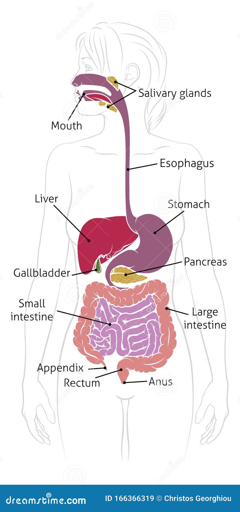
Human Digestive System Woman Anatomy Diagram Stock Vector - Illustration of gastroenterology ...
Abdomen and digestive system anatomy: diagrams labeled - IMAIOS Colon: three diagrams of the face of the colon and rectum, with or without the mesentery of the colon and taenia coli as well as a detailed illustration of the right colonic flexure. Large intestine

Human Gastrointestinal Digestive System And Labels Stock Illustration - Download Image Now - iStock
Sigmoid colon - Definition, Anatomy and Function | Kenhub Sigmoid colon - ventral view The gastrointestinal system is divided into the foregut, midgut and hindgut. The foregut stretches from the oesophagus to the major duodenal papilla, the midgut from the major duodenal papilla to two thirds of the transverse colon, and the hindgut from this point to the pectinate line of the rectum. Neurovasculature
Colorectal Cancer : Definition / West and East Medicines

Digestive System Human · Free vector graphic on Pixabay

CS100 - Spreadsheet Seminar: Components of a Spreadsheet

Reproductive Health and Fetal: Impaired function of the colon is an outline can be caused by ...
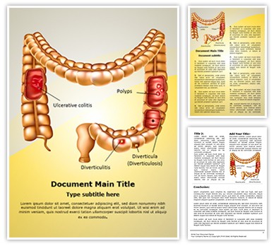
Digestive Colon Pathologies Editable Word Template and Design

Colon (anatomy) - New World Encyclopedia
Colon Problems
How The Colon Works | Colon Cleanse Recipes | Homemade Colon Cleanse

Oral Cavity Of Fetal - Human Anatomy - GUWS Medical
Post a Comment for "41 colon diagram with labels"